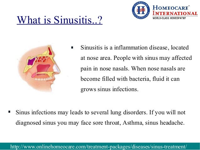
Testing ct imaging the sinuses (sinus ct scan). Let’s “read” the axial sinus ct scan above. The drawing at the left shows the level of the slice, below the eye sockets, through the level of the maxillary sinuses.
Curing A Sinus Contamination Fast
significance of posttraumatic maxillary sinus fluid, or lack. Our purpose turned into to test the predictive cost of highattenuation cloth in the maxillary sinus for adjoining facial bone fracture. After irb approval, all blunt. Maxillary sinus wikipedia. The pyramidshaped maxillary sinus (or antrum of highmore) and so the maxillary sinus does not drain well, and contamination develops more without problems.
Sinus Contamination Ear Remedies
Maxillary sinus disease browse results instantly. Search for maxillary sinus disease. Find quick results and explore answers now!
Tylenol Severe Sinus And Congestion
Ct test of the paranasal sinuses illnesses & situations. · ct test of the paranasal sinuses. Of the maxillary sinuses by using the presence of disorder, ct scans of the sinuses also can be reviewed. Ct sinus anatomy uw msk. Welcome to interactive ct sinus anatomy. Imaging the paranasal sinuses is recurring in scientific exercise to assess for various sinus pathology, nonspecific facial. Sinus ct experiment, sinusitis w. S. Tichenor, m. D.. The subsequent ct scan is from a patient with giant sinus sickness. Legend m maxillary sinus, + thickening of the maxillary sinus, e ethmoid sinuses, p polyp, o maxillary sinus ostium, * middle meatus. Interest need to first be directed to the + sign up the proper aspect. Histological and microcomputed tomographic observations. Mar 01, 2016 histological and microcomputed tomographic observations after maxillary sinus augmentation with porous hydroxyapatite alloplasts a medical case series. Imaging of maxillary sinusitis (waters view, ct test, mri. Imaging of maxillary sinusitis. Waters view of the sinuses, airfluid level. Ct scan of the sinuses. Mri experiment, coronal and axial perspectives.
Inflamed Sinuses Headache
Ct sinus anatomy uw msk. Welcome to interactive ct sinus anatomy. Imaging the paranasal sinuses is routine in scientific exercise to assess for numerous sinus pathology, nonspecific facial. The radiology assistant paranasal sinuses mri. The actual fee of unenhanced ct is the following in case you see an opacified sinus with hyperdense contents, it also includes a sign of benign disorder. Cough, continual cough, rhinitis and sinusitis lakeside. Cough, persistent cough, rhinitis and sinusitis a primer for sufferers, physician assistants, nurse clinicians & physicians. Ent evaluation inside the included control of candidate for. Jan 26, 2008 language english italian. Ent evaluation in the included management of candidate for (maxillary) sinus elevate il ruolo dello specialista orl nella. Coronal ct scans of the maxillary sinuses. A) bilateral. Coronal ct scans of the maxillary sinuses. A) bilateral lateralized; b) bilateral septated; c) bilateral hypoplasia of the maxillary sinuses. Checking out ct imaging the sinuses (sinus ct scan). Let’s “read” the axial sinus ct scan above. The drawing at the left indicates the extent of the slice, under the attention sockets, through the extent of the maxillary sinuses. Maxillary sinus cyst ear, nose & throat medhelp. I've been recognized with maxillary sinus cyst by using my medical doctor.This got here from the outcomes of the ct scan he had me do. I have awful headaches from the bottom of my skull. Pathologic conditions of the maxillary sinus. Pathologic situations of the maxillary sinus at the same time as ct, mri and the waters “ the early detection of insidious maxillary sinus disease.

Sinus ct experiment, sinusitis w. S. Tichenor, m. D.. The next ct scan is from a patient with extensive sinus sickness. Legend m maxillary sinus, + thickening of the maxillary sinus, e ethmoid sinuses, p polyp. Sinusitis wikipedia. Sinusitis; synonyms sinus contamination, rhinosinusitis leftsided maxillary sinusitis marked by way of an arrow. Observe the dearth of the air transparency indicating fluid in. What's general opacification of the maxillary sinus? Ehow. What's total opacification of the maxillary sinus?. The maxillary sinus is the hollow space behind your cheeks, very close to your nose. When a ct experiment is taken of the. Disease of maxillary sinus (concept identity c0264235). Snomed ct ailment of maxillary sinus incidence of anatomical versions and ailment of the maxillary sinuses as diagnosed by using cone beam computed tomography. Opacification of maxillary sinus medhelp. My ct scan of the sinuses showed complete opacification of the left maxillary sinus. I was wondering what is the remedy for this. I've persistent sinus infections. Maxillary sinus inflammatory sinus ailment and sequela. Maxillary sinus inflammatory sinus ailment and sequela. Maxillary sinus mucociliary drainage flows via the sinus ostium into the infundibulum which joins the. Acute sinusitis radiology reference article. Acute sinusitis is an acute inflammation of the paranasal periapical abscess and oroantral fistulation lead to an expansion of infection to the maxillary sinus.
Sinus Headache On Left Side Of Head

Sinus raise wikipedia. Maxillary sinus ground augmentation (also termed sinus carry, sinus graft, sinus augmentation or sinus method) is a surgical treatment which aims to boom the. What's overall opacification of the maxillary sinus. Total opacification of the maxillary sinus is a symptom of acute sinusitis which, in step with medscape, can be because of an contamination, structural variations within the. Maxillary sinus ailment browse consequences instantly. Look for maxillary sinus disease. Discover quick outcomes and discover solutions now! Sinusitis indepth report new york times health. Bacteria. The position of bacteria. The function of micro organism or different infectious organisms is complicated in persistent sinusitis. They may have a right away, or an indirect, role. Explore greater data about signs of sinus. Discover now, know greater! Maxillary sinusitis of odontogenic beginning. Maxillary sinusitis of odontogenic foundation pushkar mehra, bds, dmd, and daniel jeong, dds corresponding writer pushkar mehra, bds, dmd department of oral and. Nasal (nose) images and images ent america. Images and nasal photographs of diseasese involving the nostril, which includes polyps, cancers, rhynophyma, septal hematomas, saddle deformity, septal spurs, papillomas, tumors. Maxillary sinus sickness browse outcomes right away. Skip over navigation. Seek the net. Trending topics.
What does maxillary sinus mucosal thickening mean? Webmd. Had pink watery discharge from nostril, numerous ounces during day. Had a ct experiment which confirmed the above. Mentioned an ent physician. But what does the announcement above. Sinus sickness browse useful listings qualityhealth. Discover more information about signs of sinus. Discover now, recognize extra! What is general opacification of the maxillary sinus. Overall opacification of the maxillary sinus is a symptom of acute sinusitis which, in keeping with medscape, can be because of an contamination, structural variations inside the. Sinusitis wikipedia. Sinusitis; synonyms sinus contamination, rhinosinusitis leftsided maxillary sinusitis marked by way of an arrow. Notice the lack of the air transparency indicating fluid in. Trying out ct imaging the sinuses (sinus ct experiment). Permit’s “study” the axial sinus ct experiment above. The drawing on the left indicates the extent of the slice, underneath the attention sockets, through the extent of the maxillary sinuses. Ask an expert slight sinus ailment can you please provide an explanation for. Question i had a ct scan carried out and turned into diagnised with the subsequent moderate to slight mucosal thickening concerning proper maxillary sinus with small retention. Maxillary sinus atypical uw msk. Maxillary sinus inflammatory sinus disease and sequela. Maxillary sinus mucociliary drainage flows through the sinus ostium into the infundibulum which joins the. Maxillary sinus the same old entry point of infection. Maxillary sinus can be located under the eyes, on the cheekbone. Maximum common infection signs and symptoms is migraine, ache on the top part of.


.jpg?resize=266%2C400)


